Ultimate Guide to the Registered Physician in Vascular Interpretation (RPVI) Exam
In modern clinical practice, noninvasive vascular ultrasound has become the diagnostic cornerstone for assessing circulatory disorders. Accurate interpretation of these complex imaging studies directly dictates patient management, surgical planning, and clinical outcomes. For specialists in vascular surgery, cardiology, radiology, and neurology, validating this specific skill set requires a credential that stands above standard board examinations.
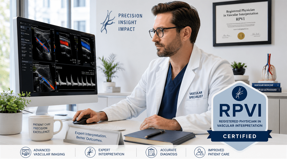
The Registered Physician in Vascular Interpretation (RPVI) certification, administered by the Alliance for Physician Certification & Advancement (APCA), represents the gold standard in noninvasive vascular laboratory interpretation. Holding this credential signifies an elite level of clinical competency and mastery over vascular hemodynamics. This comprehensive guide details the significance of the RPVI exam, career opportunities, official test domains, and how practicing with a complete set of 2120 expert-curated practice questions ensures your passing score.
The Clinical Importance of RPVI Certification
The RPVI certification is not merely an optional badge of distinction—it has increasingly become an absolute professional requirement. Notably, the Vascular Surgery Board of the American Board of Surgery (VSB-ABS) requires the RPVI credential as a mandatory prerequisite for physicians applying to sit for the Vascular Surgery Qualifying Examination.
Beyond meeting regulatory and board requirements, earning your RPVI credential proves your clinical capability to:
- Accurately grade internal carotid artery stenosis utilizing strict velocity criteria and plaque morphology assessment.
- Evaluate complex peripheral arterial disease through both duplex imaging and noninvasive physiologic testing (such as segmental pressures and waveforms).
- Identify acute and chronic deep vein thrombosis (DVT) and precisely map venous insufficiency for surgical or endovenous interventions.
- Interpret demanding visceral vascular studies, including renal artery duplex and mesenteric ischemia evaluations.
- Supervise and maintain high-quality diagnostic standards within ICAVL-accredited vascular laboratories.
Career Opportunities and Professional Value
Hospitals, multi-specialty clinics, and private imaging networks actively seek out RPVI-certified physicians to fulfill leadership and clinical oversight duties. The credential acts as an immediate validator of competence for referring physicians, insurance payors, and healthcare systems.
Key professional advantages and career paths include:
- Vascular Laboratory Medical Director: RPVI certification satisfies the demanding personnel requirements needed to direct or read in an accredited vascular lab.
- Interventional Specialist Advantage: Cardiologists and interventional radiologists can independently read and bill for noninvasive diagnostic studies, streamlining patient workflows.
- Institutional Trust & Credibility: Certified physicians serve as primary consultants for complex, ambiguous diagnostic scenarios across emergency departments, intensive care units, and outpatient clinics.
Market Value & Clinical Scope
Physicians with expert-level mastery in vascular diagnostics command significant advantages in multi-specialty medical environments. By coupling procedural skills with certified diagnostic capabilities, an RPVI-credentialed physician provides a comprehensive spectrum of care.
The clinical scope heavily impacts both institutional efficiency and physician productivity:
| Specialty Area | Core Competency Focus | Clinical Impact |
|---|---|---|
| Vascular Surgery | Pre-op planning, graft surveillance, intraoperative duplex | Mandatory Board Prerequisite |
| Cardiology / Vascular Medicine | PAD diagnosis, carotid screening, venous disease management | Expanded Lab Revenue |
| Neurology | Extracranial carotid duplex, Transcranial Doppler (TCD) | Acute Stroke Management |
RPVI Exam Blueprint and Content Domains
The Physicians' Vascular Interpretation (PVI) examination is a rigorous, computer-based test that incorporates both multiple-choice questions and rich media components (such as static ultrasound images, spectral Doppler waveforms, and video clips). Candidates must achieve a scaled passing score of 555 (on a scale from 300 to 700).
The test layout covers six core clinical domains:
- Peripheral Arterial – Duplex Imaging (20%): Interpretation of native lower/upper extremity arteries, bypass graft surveillance, endovascular stents, and dialysis access configurations.
- Peripheral Venous (20%): Evaluation of deep and superficial venous systems for thrombosis, mapping for conduit usage, and quantifying venous reflux/insufficiency.
- Cerebrovascular (15%): Analysis of the extracranial carotid/vertebral arteries and intracranial evaluation via Transcranial Doppler (TCD).
- Peripheral Arterial – Physiologic Testing (15%): Interpretation of segmental pressures, pulse volume recordings (PVR), photoplethysmography (PPG), and exercise stress testing.
- Abdominal Vascular Studies (15%): Evaluation of the abdominal aorta, iliac vessels, mesenteric arteries, and renal vasculature.
- Laboratory Technology, Physics, and Operations (15%): Concepts in fluid dynamics, Doppler physics, instrumentation artifacts, patient safety, and quality control.
Expert Preparation Strategies for Success
The RPVI exam is notoriously challenging due to its heavy emphasis on raw image interpretation, spectral waveform analysis, and underlying physics concepts. Passive reading of text is insufficient. Use these target strategies:
1. Master Diagnostic Velocity Criteria
Memorize the standardized consensus criteria for grading stenosis. You must be able to instantly calculate ratios and analyze peak systolic velocities (PSV) and end-diastolic velocities (EDV) from provided clinical images.
2. Don't Overlook Ultrasound Physics
A significant portion of candidates stumble on the physics component. Ensure you thoroughly understand Doppler angle correction, aliasing, color scale adjustment, and how to troubleshoot common imaging artifacts.
3. Commit to High-Volume Image Review
The absolute best way to build internal diagnostic pattern recognition is to review hundreds of clinical case vignettes. Practicing with structured, realistic question banks sharpens your eye for pathological variations.
Achieve Exam Mastery with the Complete 2120 Question Set
Given the rigorous demands of the APCA blueprint, high-yield practice is essential. At Killexams.com, we feature a complete set of 2120 high-quality exam questions and detailed rationales explicitly formulated for the RPVI certification exam.
Why our question engine is highly effective for busy clinicians:
- Rich Visual Scenarios: Features realistic data interpretation, spectral waveforms, and diagnostic challenges mimicking actual exam conditions.
- Complete Structural Alignment: All 2120 questions are meticulously mapped against the official six-domain APCA syllabus, ensuring zero blind spots.
- Clear Technical Explanations: Every problem is paired with a clear, direct breakdown detailing the underlying hemodynamic principles and diagnostic criteria.
- Flexible Study Optimization: Perfect for rapid retrieval, self-assessment, and diagnostic muscle memory training around busy clinical schedules.
Secure your professional advantage and protect your study investment. Approach the testing center with complete diagnostic confidence.